Study of opalised Dinosaur Vertebra

by Dr. M.S. Krzemnicki and Dr. Wei Zhou, first published in Facette 23 (February 2017)
Recently, the Swiss Gemmological Institute SSEF received an exceptional opalised dinosaur vertebra for study. This specimen was reportedly from Lightning Ridge (New South Wales in Australia) – a world famous source of dark opals and opalised plant and animal fossils – and was already photographed by Elizabeth Warren for her book Black Opal Fossils in 1999 (see page 88). The studied dinosaur vertebra is impressive in size (approx. 85 x 79 x 50 mm) and weight (501 grams), and consists of brownish greyish common opal (also known as ‘potch’) with no play-of-colour, partially interlayered with precious opal showing distinct and vivid blue and green play-of-colour. Its shape is dominated by a massive vertebra body with broken lobes, which are assumed to represent relics of the articular and traverse processes of the vertebra (see Figure 1). Based on the shape and size of the item, we assume that the studied specimen represents a distal member of the caudal vertebrae of a large dinosaur (Figure 2), such as the theropods, a saurischian suborder, which comprises the largest land-living carnivores such as the Tyrannosaurus rex (see also Warren 1999).

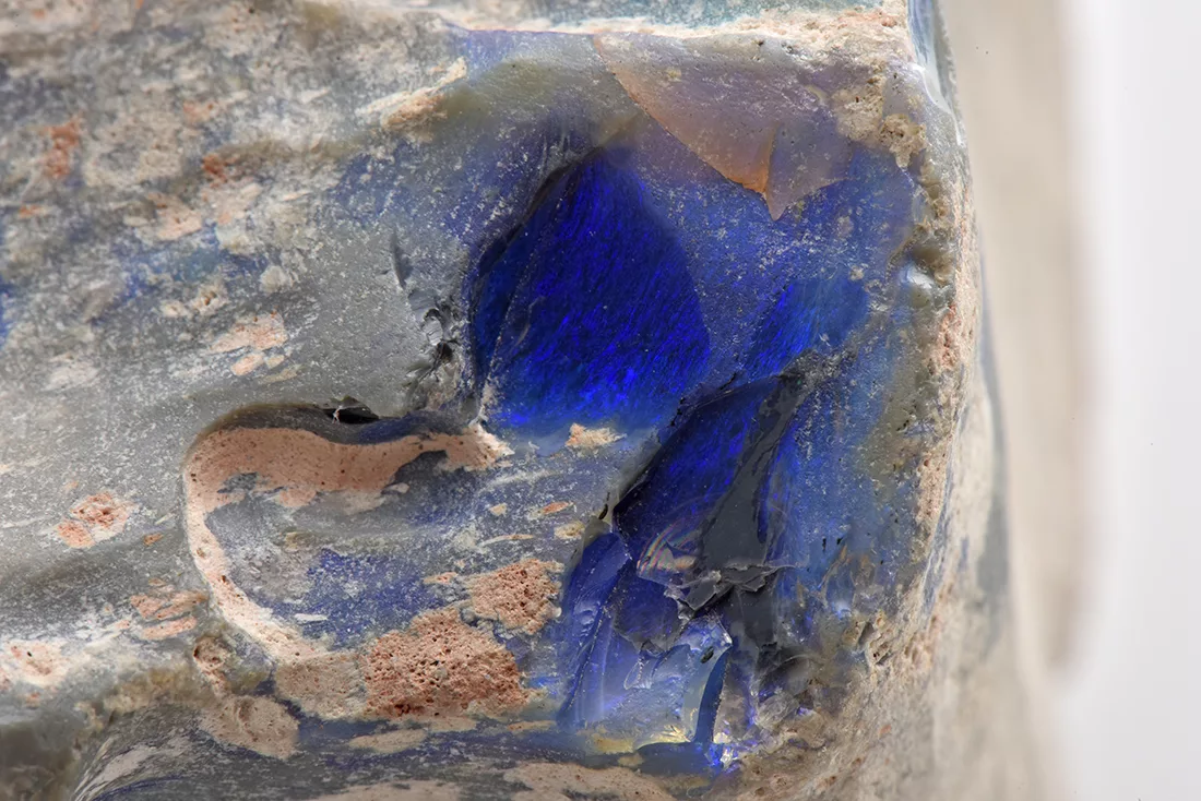
A close-up study revealed lateral linear striations at the side and a complex texture of opalised pores (probably representing the original Substantia spongiosa) at the base and top of the vertebra (Figure 3). The opalised part is best seen in a chipped off part at the side of the vertebra. It is characterised by a vivid play-of-colour, dominated by blue (Figure 4). Under ultraviolet illumination, the opalised vertebra shows patchy bluish-white fluorescence, followed by a distinct phosphorescence after switching off the UV lamp, an effect well known for opals from Australia (Gaillou et al., 2008). Due to the sample size and unpolished rough surface, only a qualitative chemical X-ray fluorescence analysis (ED-XRF) could be performed. Apart from silicon, the main constituent of opal (SiO2 x nH2O), it revealed minor amounts of calcium, potassium, iron, titanium, and traces of manganese, nickel, copper, and zinc, well known for opals from Australia (Gaillou et al., 2008).
To conclude, this opalised dinosaur vertebra is a very interesting specimen for study, as it provides not only an insight into the process of silicification of an ancient fossil into a gemmy opal with beautiful play-of-colour, but mostly also as it is a remaining trace of the fauna of Northeastern Australia in the Mesozoic.