Neutron and X-Ray Tomography of Emeralds

by Dr. Michael S. Krzemnicki, first published in Facette 23 (February 2017)
Since two years, the SSEF has been working with the Laboratory for Neutron Scattering and Imaging at the Paul Scherrer Institute (PSI) using their ICON beamline (cold neutron imaging) at the SINQ spallation source. The aim of these research projects in collaboration with Dr. Lehmann and Dr. Mannes is to analyse the internal structure of pearls and gems with neutron radiography, darkfield imaging, and tomography.
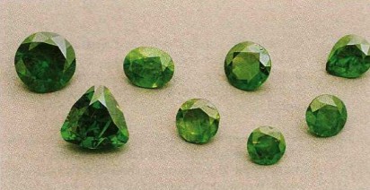
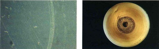
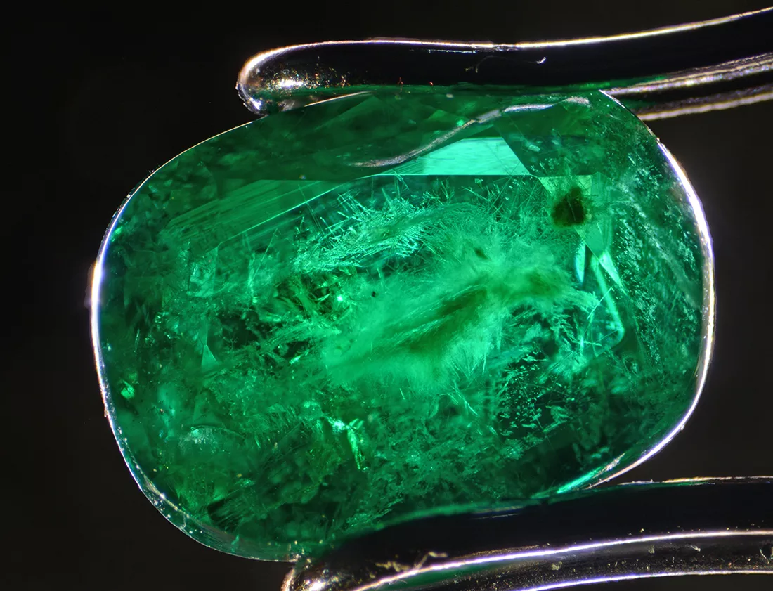
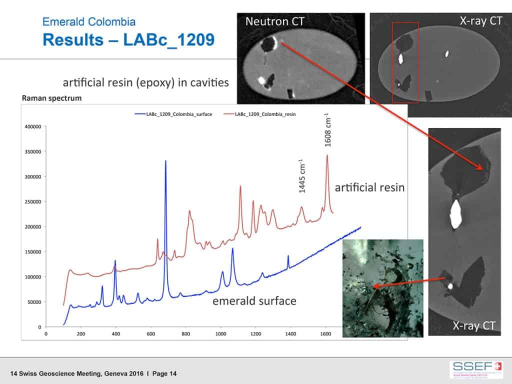
In 2016, we focused our research at PSI on the 3D-visualisation of inclusions and fissure fillings in a number of selected emeralds, notably from Colombia, Brazil, and Sandawana in Zimbabwe (Figure 1). These results were then compared to high-resolution X-ray microtomographical analyses of the same specimens.
What makes neutron imaging interesting, is that neutron interaction with atoms is not influenced by their electron cloud. This is very much in contrast to X-rays, for which the interaction (attenuation) is closely correlated to the number of electrons (and atomic number Z) of an element. Therefore, neutrons can penetrate deeply into matter which is strongly absorbing for X-rays. As such, neutron imaging may reveal important complementary information of the internal structure of materials. This is especially the case in hydrogenous materials (i.e. containing hydrogen, e.g. organics), which induce strong neutron scattering reactions.
The preliminary results of our study revealed that it is possible to visualise the complex pattern of inclusions and fissures filled with organic fillers, e.g. oil or resin in emeralds with reasonable resolution and get a 3D insight into their orientations.
Although a thorough microscopic observation assisted with FTIR and microraman spectroscopy is in most cases sufficient for the characterisation of internal features and the identification of fissure filling, this new approach will open up further possibilities in gemstone research (Mannes et al. 2016, Krzemnicki et al. 2016), such as the characterisation of ‘Trapiche’ growth patterns in gemstones, jadeite textures and impregnation, and pearl identification.
Want to learn more about emeralds?
Sign up for our free online course: Introduction to emeralds